
In Assistant Professor of Chemistry Erik Rodriguez’s lab, fluorescent proteins are lighting the way toward discoveries that could lead to, for example, breakthroughs in cancer drugs, diabetes treatments and even unlocking the brain’s secret processes for learning and memory.
But when he takes a break from pushing the boundaries of science, Rodriguez shares with students another use for these fluorescent microbes: works of art.
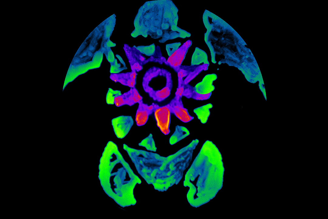
During downtime between experiments, Rodriguez has dabbled in drawing portraits of cyan turtles and tangerine unicorns. His canvas is a Petri dish. And his paint—made from bacteria expressing fluorescent proteins—is actually alive.
“It’s a fun way to break up the monotony of the lab, when, for example, you’re waiting to use a piece of equipment. It gives our students a feel for how science and art overlap,” said Rodriguez, whose fluorescent protein turtle painting was reproduced in Chemical & Engineering News (C&EN), a leading chemistry trade journal. “But, of course, this isn’t the most important use of these proteins.”
Indeed, since their discovery in the 1960s, fluorescent proteins have revolutionized biology, enabling scientists to see previously invisible aspects of cellular behavior. And Rodriguez, who recently became a member of the GW Cancer Center, has been on the forefront of many fluorescent protein milestones. He worked with 2008 Nobel Prize-winning chemist Roger Tsien, who first manipulated proteins to produce a glowing fluorescent palette that allows scientists to observe activity within cells. Rodriguez himself evolved a bright far-red fluorescent protein that allows for deeper imaging through tissue. “These proteins give us a powerful toolkit for the visualization of structural organization and dynamic processes in living cells and organisms,” he said. “We can now see pretty much any process inside the cell.”
Capturing the Glow
Fluorescent proteins first came to the science world’s attention through the North American jellyfish, Aequorea victoria. The sea creature gives off a green glow. That's because because the jellyfish, along with other species from mushrooms to fireflies, exhibits a phenomenon called bioluminescence, the ability for organisms to produce their own chemical, from which a protein then converts the chemical energy into a photon of light. The jellyfish transfers this blue light to a fluorescent protein that emits a green hue.
Scientists isolated the original green fluorescent protein (GFP) and attached the harmless fluorescent tags to selected proteins inside the cells of living creatures. When the proteins are exposed to light, researchers can track their activity, from watching neurons in the brain fire to following the path of a metastasizing cancer cell.
Eventually the jellyfish-derived GFP was engineered to push the spectrum of fluorescent proteins into blue, cyan and yellow mutations. Each glowed with different colors of light and allowed researchers to tag and study different molecules at the same time within a single cell. Red proteins, originally found in coral, have expanded the color range even further toward orange, red and far-red—spectral regions that allow for deeper molecular imaging. Typically, the farther scientists progress towards the red spectrum of light, the dimmer the fluorescence for jellyfish- and coral-derived fluorescent proteins, leading to less sensitivity for imaging. But Rodriguez’s far-red protein shines as brightly as the original GFP. These proteins have an array of potential uses—including individualizing cancer therapy, tracking the production of insulin-producing pancreas cells for new diabetes treatments and imaging calcium in the brain that may hold the answers to how humans learn and maintain memories.

Artwork by Nadine Lo, a senior assistant in Rodriguez’s lab, using fluorescent dye left over from an experiment.
Protein Painting
Rodriguez first learned to paint bacteria from his mentor Tsien. The technique involves genetically modifying bacteria to express the fluorescent proteins. Using an ordinary paint brush, Rodriguez paints an image with the bacteria on a dish. Initially, he is painting blind; the artwork only appears when the bacteria grows overnight. The light absorbed by the fluorescent proteins is seen by the eye, but the brilliant fluorescence shines in the dark with light illumination. The different fluorescent wavelengths are captured by using different filters that pass certain wavelengths of light. The images are overlaid to allow observers to see or photograph the microbial portraits.
Fluorescent protein painting is more than a chemistry lab pastime. High schools have used the technique to engage students in science. Some artists have even exhibited fluorescent portraits. Most controversially, Chicago artist Eduardo Kac unveiled a display in 2000 called “GFP Bunny,” featuring a glowing green rabbit genetically engineered with fluorescent proteins by French scientists. Rodriguez says his art is designed to illuminate students’ interests—while bringing a touch of spontaneity and art appreciation to slow lab periods when they wait for incubations or for cells to grow. Nadine Lo, a senior undergraduate assistant in Rodriguez’s lab, published her own fluorescent multi-colored dye drawing of an Andy Warhol-like eye in C&EN which she created in Rodriguez’s class.
Rodriguez’s art projects aren’t limited to painting. Two of his short films showing fluorescent proteins traveling through cells were named national winners in the 2017 Wiki Science Competition and will move on to the international competition. Meanwhile, he has no plans to trade in his microscope for a paint brush. He’s currently evolving new proteins with unique properties that continue to expand the scope of imaging technology. “The art is fun,” he said, “but our real challenge is taking [the science] as far as we can go.”